Electron microscope
An electron microscope is an electron-optical instrument in which a beam of electrons is used to produce an enlarged image of a minute object on a fluorescent screen or photographic plate. Electron microscopy is used when items or features are too small to be imaged by light. In this case, the image is created by the bending/reflection of an electron beam rather. It can magnify very small details with high resolving magnifying at levels up to 500,000 times.
Transmission electron microscope
The transmission electron microscope is much like the light microscope, but generates in image by sending an electron beam through a very thin slice of the specimen. The resolution limit is around 0.05 nanometer.
TEM gallery
Microtubule crosssection
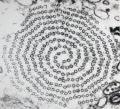
Ribosomal RNA transcription.
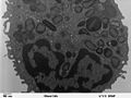
A human leukocyte (white blood cell) of the type Eosinophil.
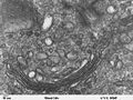
Golgi apparatus in a human leukocyte.
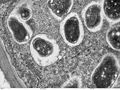
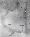
Cross section though a soybean root nodule.


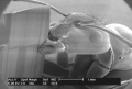
Scanning electron microscope

The scanning electron microscope is used to produce images with a characteristic three-dimensional quality. The method is for determining the surface structure of a solid by measuring the angle and energies of electrons scattered by the atoms on the surface of a sample.
SEM gallery

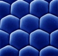
Hexagonal snow flake.

A motile spirochaete bacterium
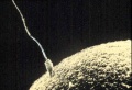
Fertilization (Sperm/Ovum)
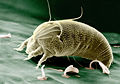
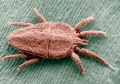
The Rust Mite (Aceria anthocoptes)
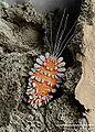
The Peacock Mite (Tuckerella sp.)
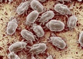
The Cheese Mite (Tyrophagus putrescentiae)
The Flat Mite (Brevipalpus phoenicis)
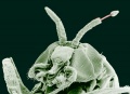
Ant head
Adult Black Fly (Simulium yahense) with parasite.
Bread mold (Penicillium chrysogenum) produces the penicillin antibiotic.
Carpenter Ant.

Salmonella bacteria (red) invading cultured human cells.
Ommatidia of eye of Antarctic krill
Compound eye of the Antarctic krill Euphausia superba.
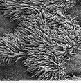
Cilia on lung trachea epithelium.
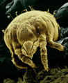
Yellow mite (Lorryia formosa), commonly found on citrus plants.
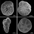
SEM micrographs of four benthic foraminiferans.
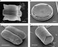
Scanning Electron Micrographs of Diatoms.

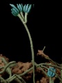
pollen from Ipomea purpurea (Heavenly blue morning glory).
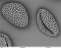
Lilium auratum pollen.
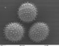
Sildalcea malviflora (prairie hollyhock) pollen.
A nerve ending broken open to reveal vesicles (orange and blue) containing neurotransmitters (chemicals used to pass messages in the nervous system).
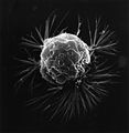
A Metastasizing Breast Cancer Cell
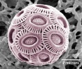
Emiliania huxleyi, a coccolithophore.
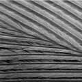
Structure of a feather.
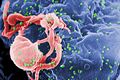
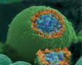
HIV-1 budding from cultured lymphocyte. Multiple round bumps on cell surface represent sites of assembly and budding of virions.

Fairy shrimp nauplius larva (about 7 hours old).
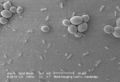
Escherichia coli (little forms) & Saccharomyces cerevisiae (big forms)
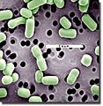
Lactobacillus brevis bacteria, which are involved with lactic acid fermentation.

Streptococcus suis bacteria. A common pathogen of pigs.

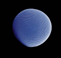
Algae (Gephyrocapsa Oceanica)
![Pollen from a variety of common plants: sunflower (Helianthus annuus), morning glory (Ipomea purpurea), hollyhock (Sildalcea malviflora), lily (Lilium auratum), primrose (Oenothera fruticosa) and castor bean (Ricinus communis).]]](https://creationwiki.org/pool/images/thumb/6/61/Microscopic_Pollen.jpg/120px-Microscopic_Pollen.jpg)
Pollen from a variety of common plants: sunflower (Helianthus annuus), morning glory (Ipomea purpurea), hollyhock (Sildalcea malviflora), lily (Lilium auratum), primrose (Oenothera fruticosa) and castor bean (Ricinus communis).]]

Water bear
(Hypsibius dujardini)
Phylum: Tardigrada


![Pollen from a variety of common plants: sunflower (Helianthus annuus), morning glory (Ipomea purpurea), hollyhock (Sildalcea malviflora), lily (Lilium auratum), primrose (Oenothera fruticosa) and castor bean (Ricinus communis).]]](/File:Microscopic_Pollen.jpg)


References
External links
Electron micrographs sources
- Dartmouth Electron Microscope Facility Publishable images by Dartmouth College
- Beltsville Electron Microscopy Unit Publishable images by Beltsville Agricultural Research Unit (USDA)
- National Center for Electron Microscopy by the Lawrence Berkeley National Laboratory
| |||||||||||||||||

