Microsporidia
| Microsporidia |
|---|

|
| Scientific Classification |
| Classes |
|
Microsporidia is a taxonomic phyla of parasitic fungi. The fungi uses a unique method of injecting their own cytoplasm into a host and then use the host to reproduce and make spores. The spores can change depending on the target host and environment, the spore stage is also the easiest way to identify separate species in this phylum. Humans can be infected with Microsporidia too, but they are mostly found in people with AIDS, and the spread of Micosporidia can be reduced by eating disinfected food and water.
Anatomy
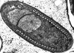
The most important and prevalent form taken during the microsporidian life cycle is that of a highly specialized single cell called the spore. When in this form the parasite is most easily recognizable, and individual species are identifiable. This is also the only form which is viable when the parasite is not infecting a living host. Spore morphology varies according to species, ranging in size from 1 - 40 microns. They are often found in the shape of an ovoid, rod, spherical, or crescent shape. The spore has walls surrounding a normal membrane, which contains the sporoplasm. This sporoplasm, which is the infectious material of the spore, contains one or two closely aligned nuclei, ribosome-enriched cytoplasm, and organelles used for infection. The most prominent among these organelles is the polar tube, located at the apex of the spore and affixed to the spore by way of an anchoring disk. From this anchoring disk the polar tube extends to the posterior end of the spore, where it comes into contact with another infection organelle, the posterior vacuole.[1]
Reproduction
The life cycle of the cell can include sexual or asexual reproduction, or both. Microsporidia with complex life cycles can have multiple hosts, and they can have 3 different spore types. Others, with simple life cycles, have very a broad tolerance and are capable of infecting nearly any type of eukaryotic cell.The spores of Microsporidia can survive for extended periods of time in the environment on their own.
The spore has a tube that is extended by a process called eversion. It turns inside-out with the dense glycoprotein core becoming an outer protective layer. The end of the tube punctures through the cell membrane of the host. The tube serves as a pipe through which the infectious sporoplasm is pumped into the host cell in 15 to 500 milliseconds. The entire process of infecting cells is completed in less than two seconds. Or, the spore may be internalized by phagocytosis (engulfing of a solid particle to form an internal vesicle)
The sporoplasm is made of the nuclei and the cytoplasm around it. In Microsporidia, ribosomes are a large component of the sporoplasm. These ribosomes promote a very high rate of protein synthesis during the first part of the infective cycle. Inside the host cell, the nuclear material in the sporoplasm replicates quickly, either inside the host cytoplasm or inside a parasite-made vacuole.
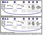
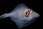
Encephalitozoon developmental stages. Although there is considerable variation, a typical microsporidian may replicate by merogony for some initial period, once it is inside the host cell. During this period, in some cases, the nuclei may proliferate with or without division into individual cells. Within the first 24-48 hours after the sporoplasm has reached the host cell, several rounds of division have occurred. When the spores completely fill the host cell cytoplasm, the cell lyses and releases the spores to the surroundings. In some systems, the microsporidian infestation may cause the development of xenomas(growths on the host)[2]
Ecology
May have several different spore types. For example, different spores may be produced on infection of primary and secondary hosts, and spores designed primarily for internal infection of additional host cells may differ from those specialized for survival in the environment.[3] Researchers studying the difference between species which infected terrestrial hosts and those which infected aquatic hosts found that overwhelmingly terrestrial species could survive in low temperatures, while aquatic species could not. This is most likely because the terrestrial parasites need to survive cooler climates during the winter so that they can initiate infections in the early spring. Exposure to extremely dry conditions yielded similar results, with some species unable to survive. Similarly, the majority of microsporidians are unable to infect hosts when exposed to overly wet or humid conditions. Exposure to sunlight kills microsporidia quickly. Some can survive for a short amount of time, but most die very quickly. Spores are spread from host to host by way of fecal matter, contaminated water and food, and inhalation. [4]
Diseases
In recent years, the US Environmental Protection Agency (EPA) has listed microsporidia in the EPA Candidate Contaminate List (CCL), deeming it an emerging water-borne pathogen needing attention. Filtering water supplies remains the best preventative strategy available. Measurement and filtration techniques for microsporidia spores remain rudimentary and underdeveloped, though the scientific community is actively trying to fix this knowledge gap. [5] There are currently no vaccines available or being explored for microsporidia infection. In humans, the most effective way to prevent the chronic diarrhea caused by microsporidia is cleanliness and decontamination of food. Although microsporidia infection in humans mostly occur in patients with compromised immune systems, the further spread of AIDS worldwide increases the need to understand and manage microsporidia for the near future. As more research is done on this class of organisms, we find their prevalence increasing in human patients. This is indeed an emerging infectious disease.
Video
Hecho por: Maria Paula Boada y Jennifer Parra Martinez
References
- ↑ [1] Microbe Wiki. Web. last midified September 14, 2015.
- ↑ . Microspridia Palaeos. Web. Month Day, Year. .Unknown Authors
- ↑ . Microspridia Palaeos. Web. Month Day, Year. .Unknown Authors
- ↑ . Microsporidia Microbe Wiki. Web. August 7, 2010. Unknown Author
- ↑ Microsporidia Stanford University. Web. accessed April 22, 2016. Unknown Author


