Atypical tarantula
| Atypical tarantula |
|---|

|
| Scientific Classification |
|
| Genera |
| Purse web |

|
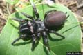
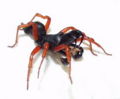
Atypical tarantulas are predatory spiders. Many spiders hunt by building webs to trap insects; these webs are made of spider silk extruded from spinnerets on the end of the abdomen, a thin, strong protein strand extruded by the spider. All spiders produce silk, even those which do not spin elaborate traps with them. Silk can be used to aid in climbing, forming smooth walls for burrows, cocooning prey, and for many other applications. Atypical tarantulas are also called purseweb spiders and consist of only three genera (genus). In the United States these are Sphodros and Atypus, and in Europe, Asia and Africa only Atypus. Atypus lives in a silken tube parallel to the surface of the ground, while Sphodros usually props its tubes against a tree trunk. The females generally do not leave their silken tubes, but catch insects that crawl on the tube by biting the prey through the silk. [1]
Gallery
purse web spider(Atypus affinis) Genera: Atypus
atypical tarantula(Sphodros rufipes) Genera: Sphodros

Distribution of Atypidae spiders around the world


References
