Heart's Electrical System
The heart begins beating at four weeks after conception and does not stop until death. [1]. The heart starts its mission since you are an embryo (21 days after start of pregnancy) to pumping blood to the entire body. Embryo heart is created before brain, and it starts to pulse from its first day till death. Research [2] by Institute of Heartmath has shown that the heart communicates to the brain in four major ways:
- neurologically (through the transmission of nerve impulses),
- biochemically (via hormones and neurotransmitters),
- biophysically (through pressure waves) and
- energetically (through electromagnetic field interactions),
Electrical stimulus is responsible for movement or muscular contraction. Heart [3] has its own internal electrical system that controls the rate and rhythm of heartbeat. With each heartbeat, an electrical signal spreads from the top of the heart to the bottom. As the signal travels, it causes the heart to contract and pump blood. What [4] keeps the heart pumping is a built-in electrical system. An electrical impulse generator, called the “sinus node,” sends signals from the right atrium to trigger the heartbeat, like a natural pacemaker. The electrical current follows a web of pathways through the heart, causing the chambers to squeeze and release in a steady, rhythmic sequence that draws blood into the heart and pushes it out.
The electrical system of our heart also is called the “Cardiac Conduction System”. This system is made up of three main parts [5] :
- The sinoatrial (SA) node, located in the right atrium of your heart
- The atrioventricular (AV) node, located on the interatrial septum close to the tricuspid valve
- The His-Purkinje system, located along the walls of your heart's ventricles
Each heartbeat has two basic parts: diastole and systole. During diastole, the atria and ventricles of your heart relax and begin to fill with blood. At the end of diastole, your heart's atria contract (atrial systole) and pump blood into the ventricles. The atria then begin to relax, heart’s ventricles then contract (ventricular systole), pumping blood out of your heart.
Each beat of the heart is set in motion by an electrical signal from within our heart muscle. In a normal, healthy heart, each beat begins with a signal from the SA node. This is why the SA node sometimes is called your heart’s natural pacemaker. Our pulse, or heart rate, is the number of signals the SA node produces per minute.
Measurement of Heart’s Activity
For a simple understanding we may say that our heart is engine of our body it makes sense that it might work something like the engine in a car, which starts with a spark. Our heart has a sparking plug and each time electrical impulse triggers because of heart’s muscles contraction, this contraction sets the timing of the whole process. The Heart “beat” is a contraction of the heart. Each time a section of the heart contracts, it forces blood from one point to another. In each heartbeat, the atrium has to contract first. The right and left atriums actually contract at the same time. What keeps the heart to beat is a built-in electrical system. An electrical impulse generator, called the “sinus node,” sends signals from the right atrium to trigger the heartbeat, like a natural pacemaker. The SA node is a group of cells that generates electrical current. The SA node sends an electrical impulse, which travels towards the AV node. While the SA node sets the rhythm of pulse, the AV node sets the rhythm of our heart contractions.
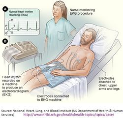
Many of the people have seen the heart test called an EKG or ECG. Generally speaking the EKG or ECG means Electrocardiogram (electro= electricity + Cardio = heart + gram = tracing) is a tracing or record of the electricity produced by contraction of the heart muscles. The painless test helps to understand how the heart works. One should not confused with ECG or EKG both are one ECG is short way to say “electrocardiogram” whereas EKG is for the German word “Electrokardiogram” both terms are used in a single sense.
ECG or EKG test that records the heart’s electrical activity is a classic example to illustrate that in reality human body produces electricity.
Pacemaker
Natural Pacemaker
The heart also has built-in “pacemakers” that are like electrical outlets. Without the electrical system, the heart would just sit there and not pump blood. The blood would not circulate and the body would not receive the oxygen and nutrients it needs. When blood flow stops to the brain, the person loses consciousness in seconds and death follows within minutes [6]. In the upper part of the right atrium of the heart is a specialized bundle of neurons known as the sinoatrial node (SA node). Acting as the heart's natural pacemaker, the SA node "fires" at regular intervals to cause the heart beat with a rhythm of about 60 to 70 beats per minute for a healthy, resting heart. The electrical impulse from the SA node triggers a sequence of electrical events in the heart to control the orderly sequence of muscle contractions that pump the blood out of the heart.
Artificial Pacemaker
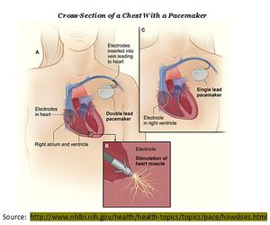
When someone needs a pacemaker, it’s usually because there’s a problem with electrical impulses, which weakens the heartbeat, causing all sorts of issues. If the heart cannot get enough blood pumping through the body, the body; and especially the brain suffers from lack of oxygen. An artificial pacemaker sends out electrical impulses to mimic the heart’s natural pacemaker[7]. An artificial pacemaker [8] consists of a battery, a computerized generator, and wires with sensors at their tips. (The sensors are called electrodes.) The battery powers the generator, and both are surrounded by a thin metal box. The wires connect the generator to the heart. A pacemaker helps monitor and control your heartbeat. The electrodes detect your heart's electrical activity and send data through the wires to the computer in the generator. Pacemakers have one to three wires that are each placed in different chambers of the heart.
- The wires in a single-chamber pacemaker usually carry pulses from the generator to the right ventricle (the lower right chamber of your heart).
- The wires in a dual-chamber pacemaker carry pulses from the generator to the right atrium (the upper right chamber of your heart) and the right ventricle. The pulses help coordinate the timing of these two chambers' contractions.
- The wires in a biventricular pacemaker carry pulses from the generator to an atrium and both ventricles. The pulses help coordinate electrical signaling between the two ventricles. This type of pacemaker also is called a cardiac resynchronization therapy (CRT) device.
Action Potential
A nerve impulse, or an action potential, is a series of electrical responses that occur in the cell. The rhythmic contractions of the heart which pump the life-giving blood occur in response to periodic electrical control pulse sequences. The natural pacemaker is a specialized bundle of nerve fibers called the sinoatrial node (SA node). Nerve cells are capable of producing electrical impulses called action potentials. Electricity is a movement of charge. In a wire the electrons move to carry the charge along the wire. In a nerve the electrical signal moves although no electrical charge actually moves along the nerve - the signal travels in the form of an action potential. The inside of nerve cells normally has a slight negative charge as a result of the activity of pumps which move charged ions predominantly out of the cell. When an electrical charge is placed near a nerve cell it causes gates in the membrane to open and allow the ions to reenter the cell and depolarize the membrane near the position where the charge is located. The depolarization causes ion channels to open in the membrane a little further along the nerve (resulting in depolarization here) and then channels open in the membrane a little further along...the chain reaction of depolarization moves along the nerve until it gets to the end where the depolarization causes an appropriate response (such as muscle contraction) [9].
Action potentials in neurons are also known as "nerve impulses" or "spikes", and the temporal sequence of action potentials generated by a neuron is called its "spike train". A neuron that emits an action potential is often said to “fire”. Action potentials are generated by special types of voltage-gated ion channels embedded in a cell’s plasma membrane. The electrical potentials (voltages) differences in the concentrations of positive and negative ions give a localized separation of charges. This charge separation is called polarization. Changes in voltage occur when some event triggers a depolarization of a membrane, and also upon the depolarization of the membrane. The depolarization and depolarization of the SA node and the other elements of the heart’s electrical system produce a strong pattern of voltage change which can be measured with electrodes on the skin.
References
- ↑ http://facts.randomhistory.com/human-heart-facts.html
- ↑ http://www.heartmath.org/free-services/articles-of-the-heart/heart-brain-interactions.html
- ↑ http://www.nhlbi.nih.gov/health/health-topics/topics/ (different topics)
- ↑ http://www.arrhythmia.org/electrical.html
- ↑ http://www.nhlbi.nih.gov/health/health-topics/topics/pace/
- ↑ Source: http://www.arrhythmia.org/electrical.html
- ↑ http://health.howstuffworks.com/human-body/systems/circulatory/heart-rhythm.htm/printable
- ↑ http://www.nhlbi.nih.gov/health/health-topics/topics/pace/howdoes.html
- ↑ Is there electricity anywhere else in the Human Body? Answer by: Cameron Millsom - http://www.newton.dep.anl.gov/askasci/bio99/bio99800.htm


