Uploads by Tian-yi
From CreationWiki
Jump to navigationJump to search
This special page shows all uploaded files.
| Date | Name | Thumbnail | Size | Description | Versions |
|---|---|---|---|---|---|
| 08:41, 16 June 2008 | Electrolytic cell.gif (file) |  |
4 KB | An electrolytic cell for leak examination. | 1 |
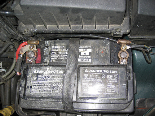
| 06:52, 16 June 2008 | Lead Storage Battery in the car.jpg (file) |  |
144 KB | 1 | |
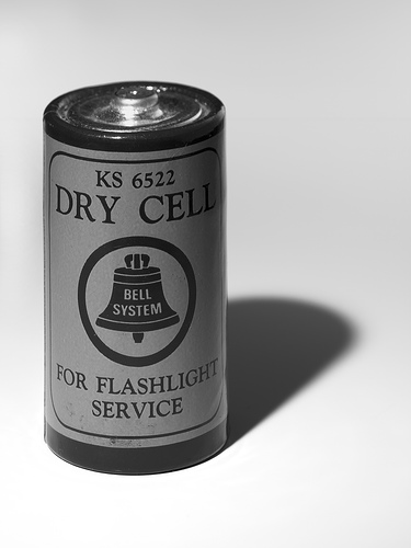
| 06:42, 16 June 2008 | Dry Cell Flashlight Battery.jpg (file) |  |
41 KB | 1 | |
| 02:00, 4 June 2008 | Stracciabraghe.jpg (file) |  |
117 KB | Smilax aspera | 1 |
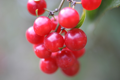
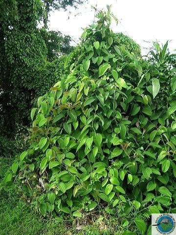
| 01:57, 4 June 2008 | Smilax australis.jpg (file) |  |
134 KB | Smilax australis | 1 |
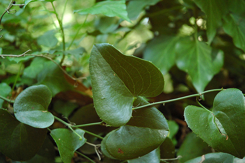
| 01:52, 4 June 2008 | Smilax aspera.jpg (file) |  |
55 KB | Smilax aspera | 1 |
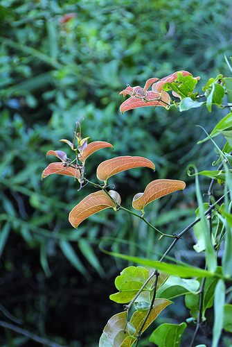
| 19:37, 30 May 2008 | Smilax bona-nox.jpg (file) |  |
88 KB | Smilax bona-nox | 1 |
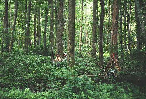
| 04:05, 25 May 2008 | Swamp Forest.jpg (file) |  |
157 KB | An 80-year old non-riverine swamp forest in the eastern part of the Great Dismal Swamp, near the headwaters of the Northwest River, City of Chesapeake. Red maple (Acer rubrum), sweetgum (Liquidambar styraciflua), bald cypress (Taxodium distichum), and wat | 1 |
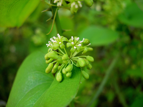
| 19:50, 9 May 2008 | Smilax setosa.jpg (file) |  |
99 KB | Smilax setosa. Singapore category:Smilacaceae | 1 |
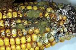
| 05:16, 29 April 2008 | Aspergillus.jpg (file) |  |
28 KB | Toxin-producing Aspergillus flavus mold is the greenish powdery growth on this ear of corn damaged by corn rootworms. | 1 |
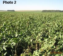
| 05:11, 29 April 2008 | Sclerotinia.jpg (file) |  |
17 KB | Sclerotinia affecting soybeans in Tandil will reduce yield by 2 percent in this field. | 1 |
| 21:49, 24 April 2008 | Western Diamondback Rattlesnake.jpg (file) |  |
83 KB | Reverted to version as of 01:59, 4 January 2008 | 3 |
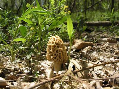
| 01:48, 24 April 2008 | Morel.jpg (file) |  |
34 KB | 1 | |
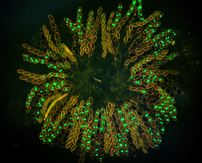
| 01:08, 24 April 2008 | Ascus.jpg (file) |  |
171 KB | Neurospora asci. The asci are from a cross of histone H1-GFP x wild type. Four of the eight ascospores in each ascus show glowing hH1-GFP nuclei. The remaining four ascospores contain non-glowing wild type nuclei. | 1 |
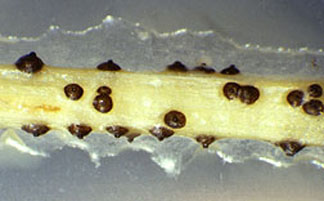
| 00:59, 24 April 2008 | Pycnidia.jpg (file) |  |
17 KB | Black pycnidia of Phomopsis developing in culture on stems of alfalfa | 2 |
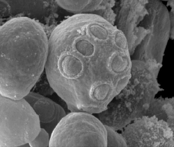
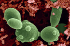
| 23:23, 23 April 2008 | Saccharomyces cells 2.jpg (file) |  |
40 KB | Scanning electrograph image of Saccharomyces cerevisiae cells grown on the International Space Station. | 1 |
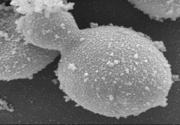
| 23:19, 23 April 2008 | Saccharomyces cells.jpg (file) |  |
41 KB | Scanning electrograph image of Saccharomyces cerevisiae cells grown on Earth. | 1 |
| 19:04, 21 April 2008 | Saccharomyces.jpg (file) |  |
40 KB | Saccharomyces boulardi (large cells) found along with bacteria in fermented fruit juice. Photo Credit: SciMAT / Photo Researchers, Inc | 1 |
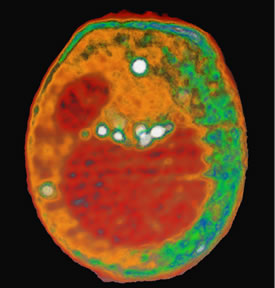
| 15:02, 21 April 2008 | Yeast cell.jpg (file) |  |
18 KB | This x-ray tomography image of a yeast cell taken at the ALS with XM-1 is an example of what could be done with the proposed XM-2. Internal organelles are color-coded according to x-ray absorption. Shown in red are the nucleus (smaller sphere) and large v | 1 |
| 14:58, 21 April 2008 | 177674main Yeast-GAP3.jpg (file) |  |
40 KB | Scanning electrograph image of Saccharomyces cerevisiae cells grown on the International Space Station. | 1 |
| 14:54, 21 April 2008 | 177672main Yeast-GAP2.jpg (file) |  |
41 KB | 1 | |
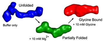
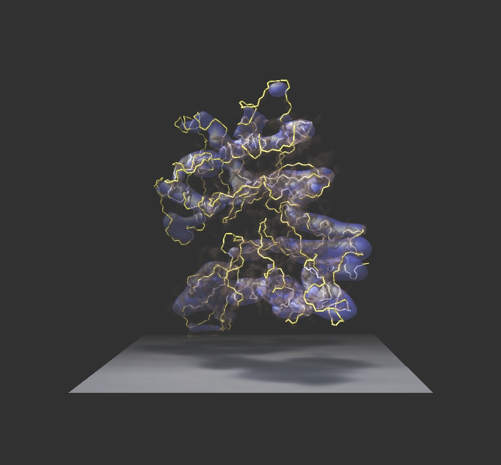
| 03:44, 1 April 2008 | Glycine binding.fig2.png (file) |  |
9 KB | Low-resolution 3-D reconstructions from SAXS measurements of a glycine binding riboswitch. Unfolded conformation in the absence of divalent counterions (blue), partially folded conformation in the presence of Mg2+ counterions (green), and fully folded con | 1 |
| 17:24, 31 March 2008 | Glycine.jpg (file) |  |
26 KB | Glycine-nitrate process produces ultrafine metal oxide powders. | 1 |
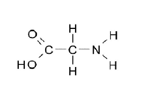
| 17:31, 20 March 2008 | Glycine.JPG (file) |  |
9 KB | 1 | |
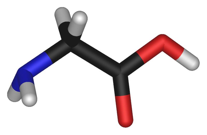
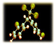
| 17:51, 12 March 2008 | Glycine-3D-sticks.JPG (file) |  |
20 KB | 1 | |
| 08:41, 8 March 2008 | Modifying plants.jpg (file) |  |
16 KB | 1 | |
| 06:18, 3 March 2008 | Biotechnology.gif (file) |  |
35 KB | Biotechnology Division (NIST Chemical Science and Technology Laboratory) | 1 |
| 22:08, 22 January 2008 | Dna replication.gif (file) |  |
41 KB | 1 | |
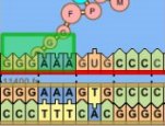
| 20:27, 22 January 2008 | DNA synthesis picture.jpg (file) |  |
7 KB | A image of DNA synthesis | 1 |
| 04:48, 19 January 2008 | Cancer tumor.jpg (file) |  |
50 KB | Cells of an ovarian cancer tumor treated with Exherin viewed through a light microscope. The vessel has ballooned and ruptured, allowing red blood cells (RBCs) to pool and escape into the tumor | 1 |
| 21:34, 15 January 2008 | DNA synthesis.jpg (file) |  |
9 KB | 1 | |
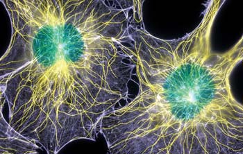
| 06:06, 20 November 2007 | Microtubule.jpg (file) |  |
24 KB | In these cells, actin filaments appear light purple, microtubules yellow, and nuclei greenish blue. | 1 |
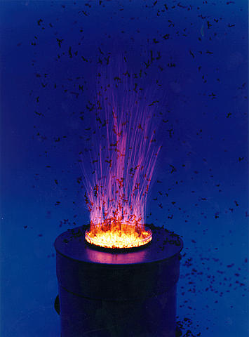
| 06:00, 20 November 2007 | Intermediate filaments.jpg (file) |  |
5 KB | This fireworks explosion of color is a dividing newt lung cell seen under a light microscope and colored using fluorescent dyes: chromosomes in blue, intermediate filaments in red, and spindle fibers (bundled microtubules assembled for cell division) in g | 1 |
| 05:34, 20 November 2007 | Cytoskeleton.jpg (file) |  |
181 KB | Actin and tubulin are both protein molecules that polymerize into fibrous structures, known as the cytoskeleton, inside cells. Both types of fibers are involved in establishing cell shape, cell movement, and moving things around inside the cells. | 1 |
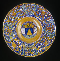
| 06:53, 19 November 2007 | Tin oxide plate.jpg (file) |  |
31 KB | A glaze containing tin oxide sealed the clay body and provided an opaque white base for decoration. | 1 |
| 06:49, 19 November 2007 | Tin oxide plste.jpg (file) |  |
31 KB | A glaze containing tin oxide sealed the clay body and provided an opaque white base for decoration. | 1 |
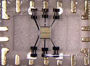
| 07:49, 17 November 2007 | Tin oxide.jpg (file) |  |
72 KB | Tin oxide microsensor used for fire detection. | 1 |
| 23:06, 26 October 2007 | Movement.jpg (file) |  |
16 KB | 1 | |
| 22:37, 26 October 2007 | Legs animation.gif (file) |  |
52 KB | Look at the flashes of red when the legs walk forward. These are the working muscles as they contract; the muscles in yellow are at rest. Rehabilitation medicine specialists use this imaging technology for gait analysis, how people walk or run. By mapping | 1 |
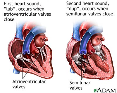
| 19:52, 25 October 2007 | Heart beat.jpg (file) |  |
35 KB | Two distinguishable sounds can be heard during the cycle of the beating heart when listened to with a stethoscope. The heart sounds are usually described as a lup-dup sound. These sounds are due to the closing of the valves of the heart. Unusual heart sou | 1 |
| 19:50, 25 October 2007 | Muscle contraction.PNG (file) |  |
190 KB | 4 | |
| 19:14, 25 October 2007 | The Structure of Skeletal muscle.PNG (file) |  |
23 KB | Each muscle cell contains many small bundles of contractile proteins, called myofibrils. These contractile proteins do the work of muscle contraction. category:Muscular system | 1 |
| 19:02, 25 October 2007 | Skeletal muscle cell.gif (file) |  |
24 KB | 1 | |
| 17:25, 12 October 2007 | Vanadinite.png (file) |  |
107 KB | Vanadium is not found in the native state, but is present in minerals such as vanadinite, PB5(VO4)3Cl. category:Vanadium | 1 |
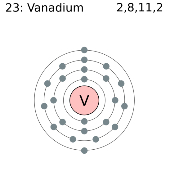
| 17:20, 12 October 2007 | Electron shell of vanadium.png (file) |  |
46 KB | 1 | |
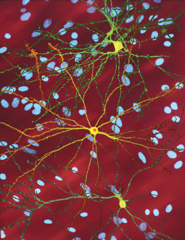
| 22:55, 4 October 2007 | Neurons.jpg (file) |  |
60 KB | A montage of three images of single striatal neurons transfected with a disease-associated version of huntingtin, the protein that causes Huntington's disease. Nuclei of untransfected neurons are seen in the background (blue). The neuron in the center (ye | 2 |
| 21:42, 3 October 2007 | Nervous system diagram.png (file) |  |
176 KB | English: A diagram of the Human Nervous system. Français : Schéma du système nerveux humain. category:nervous system | 1 |