Uploads by Felicia.file
From CreationWiki
Jump to navigationJump to search
This special page shows all uploaded files.
| Date | Name | Thumbnail | Size | Description | Versions |
|---|---|---|---|---|---|
| 02:23, 8 May 2013 | 494px-Esophagus, stomach, small intestine.jpg (file) |  |
32 KB | Line drawing showing the esophagus, stomach and small intestine. | 1 |
| 02:16, 8 May 2013 | Layers.gif (file) |  |
51 KB | Section of the human esophagus. (From a drawing by V. Horsley.) Moderately magnified. The section is transverse and from near the middle of the gullet. a. Fibrous covering. b. Divided fibers of longitudinal muscular coat. c. Transverse muscular fibers. d. | 1 |
| 02:13, 8 May 2013 | Esophagus.gif (file) |  |
75 KB | The position and relation of the esophagus in the cervical region and in the posterior mediastinum. Seen from behind | 1 |
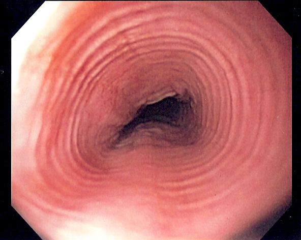
| 02:48, 25 April 2013 | Multi ring esophagus.jpg (file) |  |
80 KB | Endoscopic picture of multi-ring esophagus seen in a patient with en:eosinophilic esophagitis | 1 |
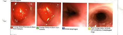
| 02:26, 25 April 2013 | Esophagus.jpg (file) |  |
8 KB | Esophagus | 1 |
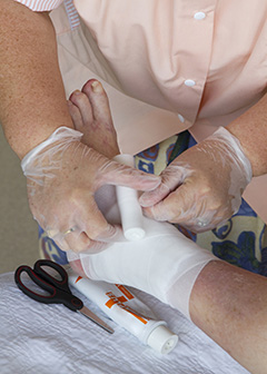
| 03:10, 27 February 2013 | Lkjsdfkjsdf.jpg (file) |  |
87 KB | Podiatrist in a consultation with a patient. | 1 |
| 01:00, 11 February 2013 | Help.jpg (file) |  |
36 KB | Patients with diabetes may develop foot problems that require the care of a podiatrist. | 1 |
| 00:52, 11 February 2013 | Injured foot.jpg (file) |  |
33 KB | Podiatrists treat common foot and ankle ailments as well as perform more complicated surgeries. | 1 |
| 00:49, 11 February 2013 | Podiatrist.jpg (file) |  |
29 KB | Podiatrists use x rays to diagnose foot and ankle problems. | 1 |
| 02:19, 16 November 2012 | Bull's eye rash.jpg (file) |  |
28 KB | Rash occurs in approximately 70-80% of infected persons1 and begins at the site of a tick bite after a delay of 3-30 days (average is about 7 days). Rash gradually expands over a period of several days, and can reach up to 12 inches (30 cm) across. Parts | 1 |
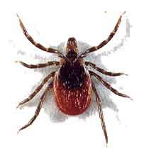
| 02:57, 30 October 2012 | DeerTick2.jpg (file) |  |
4 KB | Deer Tick | 1 |
| 02:54, 30 October 2012 | Cdc photo of lyme disease bullseye rash.jpg (file) |  |
24 KB | Rash from deer tick bite | 1 |
| 02:46, 30 October 2012 | Lyme diseas map pic.GIF (file) |  |
20 KB | Map of approximate distribution of Lyme disease | 1 |
| 19:41, 27 January 2012 | Calcium-carbonate dot structure diagram.png (file) |  |
21 KB | 1 | |
| 19:37, 27 January 2012 | Calcium carbonate letter structure.png (file) |  |
5 KB | 1 | |
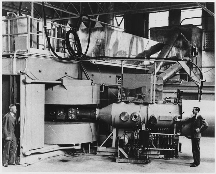
| 02:43, 18 November 2011 | 747px-Berkeley 60-inch cyclotron gif.jpg (file) |  |
87 KB | Photograph shows the 60-inch cyclotron at the University of California Lawrence Radiation Laboratory, Berkeley, in August, 1939. The machine was the most powerful atom-smasher in the world at the time. It had started operating early in the year. During th | 1 |
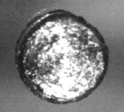
| 04:07, 4 November 2011 | Californium 2.jpg (file) |  |
19 KB | Silvery | 1 |
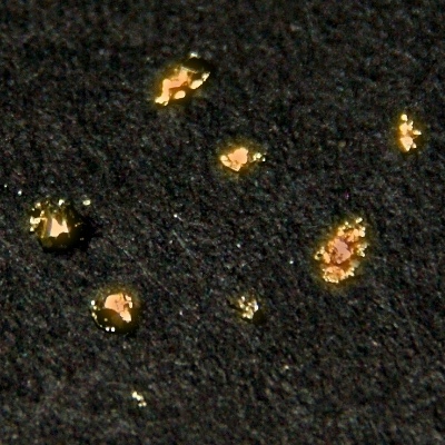
| 04:04, 4 November 2011 | Californium.jpg (file) |  |
125 KB | This is only an illustration, not californium itself. The strong α and neutron emitter produces a considerable heat. | 1 |
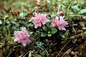
| 01:01, 8 May 2011 | 29325 large.jpg (file) |  |
19 KB | Rhododendron lapponicum (L.) Wahlenb. | 1 |
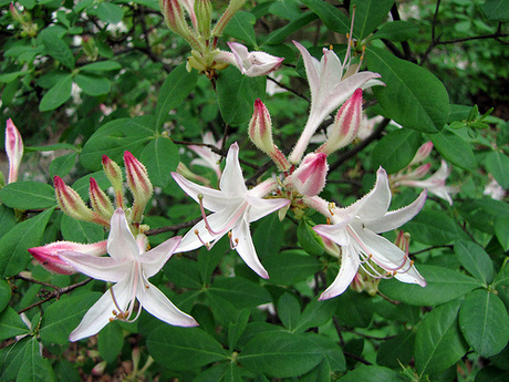
| 00:54, 8 May 2011 | Adf.jpg (file) |  |
140 KB | Rhododendron atlanticum (Ashe) Rehd. | 1 |
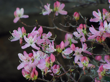
| 00:40, 8 May 2011 | 88493 large.jpg (file) |  |
121 KB | Rhododendron vaseyi Gray | 1 |
| 00:27, 8 May 2011 | 56177 large.jpg (file) |  |
77 KB | Rhododendron prinophyllum (Small) Millais | 1 |
| 03:45, 22 April 2011 | Rhododendron japonicum (Gray) Sur..jpg (file) | _Sur..jpg) |
14 KB | Rhododendron japonicum (Gray) Sur. Japanese azalea | 1 |
| 03:39, 22 April 2011 | Rhododendron oblongifolium.jpg (file) |  |
95 KB | Rhododendron oblongifolium (Small) Millais Texas azalea | 1 |
| 03:33, 22 April 2011 | Rhododendron atlanticum.jpg (file) |  |
140 KB | Rhododendron atlanticum (Ashe) Rehd. Dwarf azalea | 1 |
| 03:26, 22 April 2011 | Rhododendron molle.jpg (file) |  |
99 KB | Shrubs, 0.5–2 m tall; branches densely gray-white-pubescent, also sparsely setose when young. Petiole 2–6 mm, puberulent and ± setose; leaf blade papery, oblong to oblong-lanceolate, 5–11 × 1.5–3.5 cm; base cuneate; margin ciliate; apex obtuse a | 1 |
| 03:36, 16 February 2011 | Beluga3.jpg (file) |  |
113 KB | The beluga whale is a northern hemisphere species, ranging primarily over the Arctic Ocean and some adjoining seas, where it inhabits fjords, estuaries, and shallow water in Arctic and subarctic oceans. Belugas generally occur in shallow, coastal waters, | 1 |
| 05:09, 4 February 2011 | Baby beluga1.jpg (file) |  |
407 KB | Scientists tagging a beluga whale in Cook Inlet near Anchorage. | 1 |
| 05:02, 4 February 2011 | Belugawhale regions.jpg (file) |  |
64 KB | Beluga Whale Range Map | 1 |
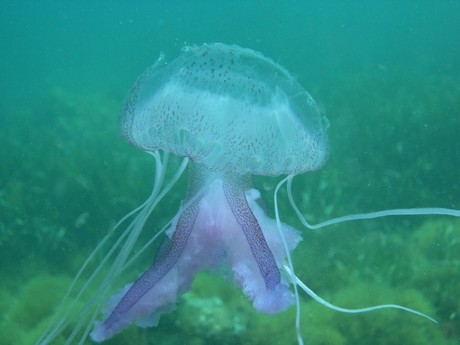
| 01:33, 10 December 2010 | Pelagia noctiluca purplestriped jellyfish2.jpg (file) |  |
169 KB | pelagia noctiluca purplestriped jellyfish | 1 |
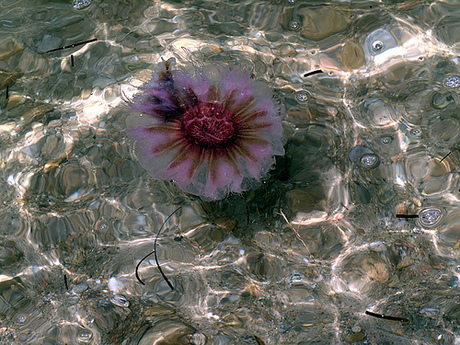
| 01:26, 10 December 2010 | Pelagia noctiluca purplestriped jellyfish.jpg (file) |  |
43 KB | Pelagia noctiluca the purplestriped jellyfish. Atlantic coast of Nova Scotia | 1 |
| 18:38, 3 December 2010 | Pelagia noctiluca 2.jpg (file) |  |
28 KB | Pelagia noctiluca (Forskål, 1775) Purplestripped jelly | 1 |
| 18:30, 3 December 2010 | Pelagia noctiluca.jpg (file) |  |
115 KB | Pelagia noctiluca (Forskål, 1775) Purplestripped jelly | 1 |