File:Lung cilia.jpg
From CreationWiki, the encyclopedia of creation science
Jump to navigationJump to search
Size of this preview: 744 × 599 pixels. Other resolution: 1,560 × 1,257 pixels.
Original file (1,560 × 1,257 pixels, file size: 724 KB, MIME type: image/jpeg)
|
This image was uploaded to the shared "Image Pool" and is usable on any CreationWiki site. |
File history
Click on a date/time to view the file as it appeared at that time.
| Date/Time | Thumbnail | Dimensions | User | Comment | |
|---|---|---|---|---|---|
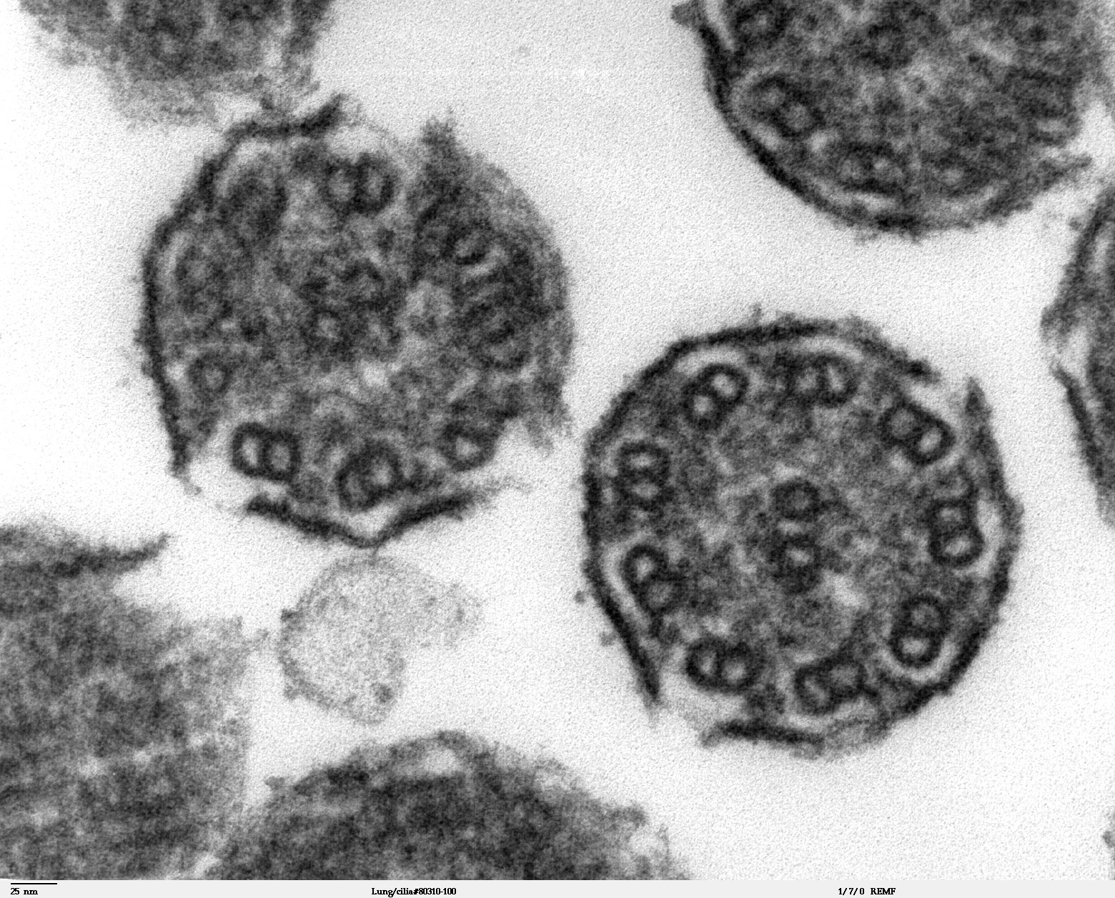
| current | 01:45, 28 November 2009 | | 1,560 × 1,257 (724 KB) | Ashcraft | High magnification of "Lung cilia#80311-50" image. This image is a thin x-section cut through cilia. The inside of the cilium contain precisely arranged microtubles, the axoneme. The axoneme has a central unit containing two single microtubules and nine p |
File usage
The following 2 pages use this file: