File:Microtubure crosssection.jpg
From CreationWiki
Jump to navigationJump to search
No higher resolution available.
Microtubure_crosssection.jpg (658 × 600 pixels, file size: 81 KB, MIME type: image/jpeg)
Summary
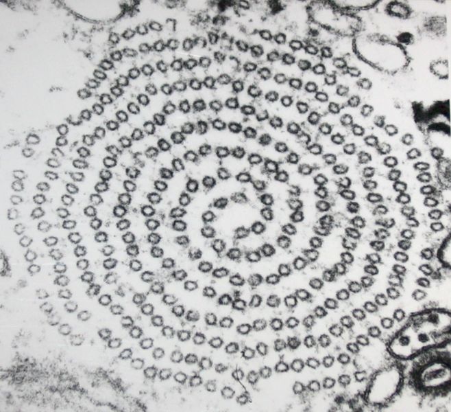
Querschnitt durch ein Axopodium (Heliozoa) als Substrukturen werden zwei spiralförmig angeordnete Mikrotubulireihen erkennbar. Transmissionselektronenmikroskopie, Vergr.: ca 65 000x Foto und Repro Michael Linnenbach
Copyright status:

|
Permission is granted to copy, distribute and/or modify this document under the terms of the GNU Free Documentation License, Version 1.2 or any later version published by the Free Software Foundation; with no Invariant Sections, no Front-Cover Texts, and no Back-Cover Texts. A copy of the license is included in the section entitled GNU Free Documentation License. | ||||||||||
This file is licensed under the Creative Commons Attribution-Share Alike 3.0 Unported license. You are free: • to share – to copy, distribute and transmit the work. • to remix – to adapt the work Under the following conditions: • attribution – You must attribute the work in the manner specified by the author or licensor (but not in any way that suggests that they endorse you or your use of the work). • share alike – If you alter, transform, or build upon this work, you may distribute the resulting work only under the same or similar license to this one. | |||||||||||
Source:
http://commons.wikimedia.org/wiki/File:Axopodium_Mikrotubuli.jpg
File history
Click on a date/time to view the file as it appeared at that time.
| Date/Time | Thumbnail | Dimensions | User | Comment | |
|---|---|---|---|---|---|
| current | 22:35, 16 March 2014 | | 658 × 600 (81 KB) | Luiz Alexandre Silva (talk | contribs) | Querschnitt durch ein Axopodium (Heliozoa) als Substrukturen werden zwei spiralförmig angeordnete Mikrotubulireihen erkennbar. Transmissionselektronenmikroskopie, Vergr.: ca 65 000x Foto und Repro [http://de.wikipedia.org/wiki/Benutzer:Michael_Linnenbach |
You cannot overwrite this file.
File usage
There are no pages that use this file.