File:Microtubule structure.jpg

Original file (650 × 608 pixels, file size: 184 KB, MIME type: image/jpeg)
Summary
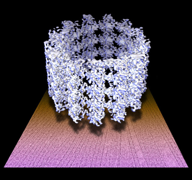
Three dimensional microtubule map floating on a raw electron micrograph.
Microtubules play important roles in eukaryotic cell morphogenesis, organelle transport, and mitosis. During mitosis, the microtubule network is restructured to form the mitotic apparatus. This requires considerable dynamics of the microtuble. Interaction between the component tubulin molecules is the key to understanding this dynamic property. In the 8Å resolution 3D map determined by cryo-EM and single particle image analysis, tubulin secondary structures, such as alpha helices, are well defined. This warrants an accurate positioning of the tubulin atomic structures into the microtubule map, and provides unambiguous information about the intermolecular interactions.
Copyright status
This image is public domain because it was first published by the Brookhaven National Laboratory.
Source
File history
Click on a date/time to view the file as it appeared at that time.
| Date/Time | Thumbnail | Dimensions | User | Comment | |
|---|---|---|---|---|---|
| current | 00:22, 15 January 2008 | | 650 × 608 (184 KB) | Ashcraft (talk | contribs) | Three dimensional microtubule map floating on a raw electron micrograph. Microtubules play important roles in eukaryotic cell morphogenesis, organelle transport, and mitosis. During mitosis, the microtubule network is restructured to form the mitotic ap |
You cannot overwrite this file.
File usage
There are no pages that use this file.