File:Lungtrachea.jpg
Lungtrachea.jpg (512 × 525 pixels, file size: 69 KB, MIME type: image/jpeg)
Краткое описание
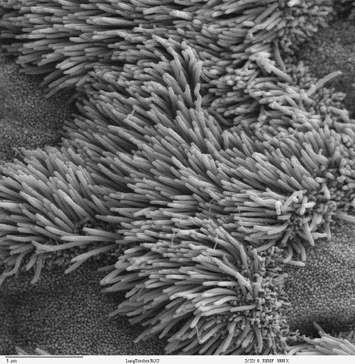
Scanning electron microscope image of lung trachea epithelium. There are both ciliated and non-ciliated cells in this epithelium. Note the difference in size between the cilia and the microvilli(on non-ciliated cell surface)
Zeiss DSM 962 SEM
Copyright status:
These images are in the public domain. Do with them what you will. If you have questions about the images or the microscopy and specimen preparation used to obtain images contact Louisa Howard . For questions regarding the research on the specimens, contact the person noted in the caption of the images.
http://remf.dartmouth.edu/imagesindex.html
Source:
http://remf.dartmouth.edu/images/mammalianLungSEM/source/12.html
File history
Click on a date/time to view the file as it appeared at that time.
| Date/Time | Thumbnail | Dimensions | User | Comment | |
|---|---|---|---|---|---|
| current | 06:21, 10 June 2010 | | 512 × 525 (69 KB) | Alexander Korolev (talk | contribs) | Scanning electron microscope image of lung trachea epithelium. There are both ciliated and non-ciliated cells in this epithelium. Note the difference in size between the cilia and the microvilli(on non-ciliated cell su |
You cannot overwrite this file.
File usage
There are no pages that use this file.