File:Classified Diatoms.png
Original file (1,400 × 1,144 pixels, file size: 951 KB, MIME type: image/png)
Summary
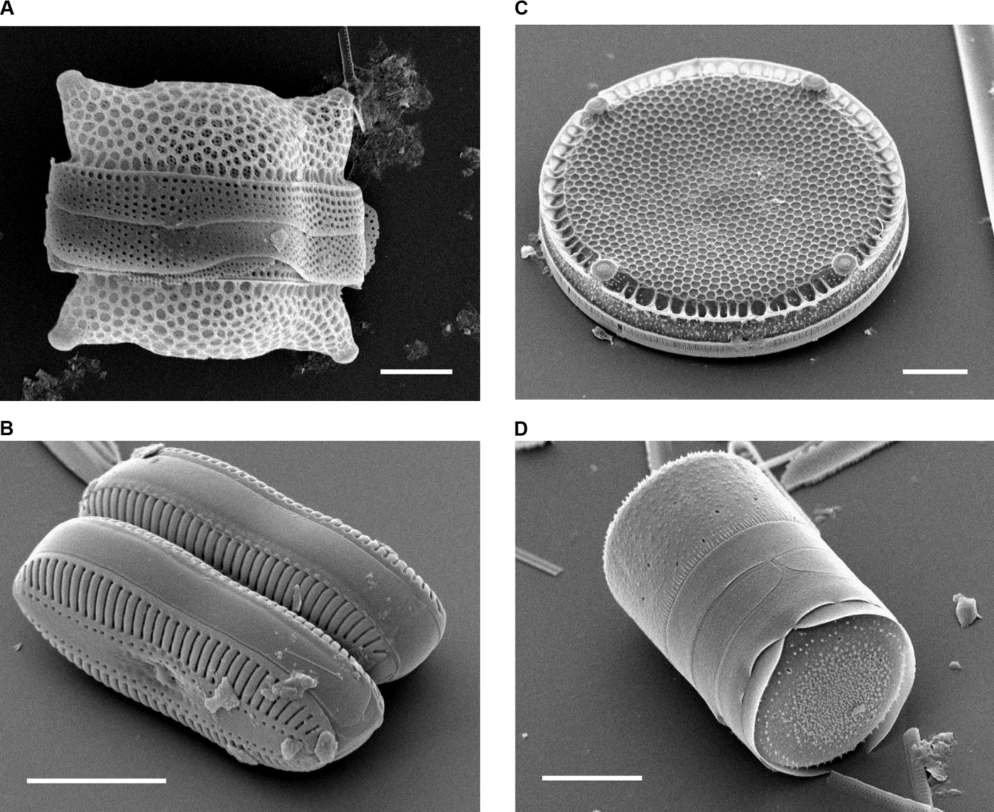
Scanning Electron Micrographs of Diatoms. (A) Biddulphia reticulata. The whole shell or frustule of a centric diatom showing valves and girdle bands (size bar = 10 micrometres). (B) Diploneis sp. This picture shows two whole pennate diatom frustules in which raphes or slits, valves, and girdle bands can be seen (size bar = 10 micrometres). (C) Eupodiscus radiatus. View of a single valve of a centric diatom (size bar = 20 micrometres) (D) Melosira varians. The frustule of a centric diatom, showing both valves and some girdle bands (size bar = 10 micrometres).
Copyright status
This image was published in a Public Library of Science journal. Their website states that the content of all PLoS journals is published under the Creative Commons Attribution.
Source
File history
Click on a date/time to view the file as it appeared at that time.
| Date/Time | Thumbnail | Dimensions | User | Comment | |
|---|---|---|---|---|---|
| current | 03:09, 19 March 2007 | | 1,400 × 1,144 (951 KB) | Dlee (talk | contribs) | Classified diatoms |
You cannot overwrite this file.
File usage
There are no pages that use this file.