File:Yeast cell.jpg
Yeast_cell.jpg (275 × 288 pixels, file size: 18 KB, MIME type: image/jpeg)
Summary
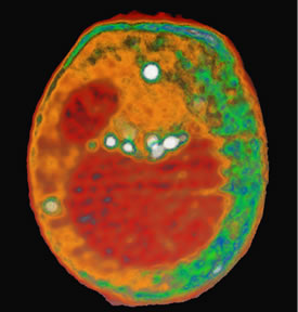
This x-ray tomography image of a yeast cell taken at the ALS with XM-1 is an example of what could be done with the proposed XM-2. Internal organelles are color-coded according to x-ray absorption. Shown in red are the nucleus (smaller sphere) and large vacuole. Lipid droplets are shown in white, and cytoplasmic structures are shown in either orange or green.
Copyright status
This picture is public domain because it's public by LBL
Source
http://www.lbl.gov/Science-Articles/Archive/assets/images/2004/Mar-30/yeast.jpg
http://www.lbl.gov/Science-Articles/Archive/ALS-x-ray-microscopy.html
File history
Click on a date/time to view the file as it appeared at that time.
| Date/Time | Thumbnail | Dimensions | User | Comment | |
|---|---|---|---|---|---|
| current | 15:02, 21 April 2008 | | 275 × 288 (18 KB) | Tian-yi (talk | contribs) | This x-ray tomography image of a yeast cell taken at the ALS with XM-1 is an example of what could be done with the proposed XM-2. Internal organelles are color-coded according to x-ray absorption. Shown in red are the nucleus (smaller sphere) and large v |
You cannot overwrite this file.
File usage
There are no pages that use this file.