File:Neurons.jpg
Neurons.jpg (362 × 470 pixels, file size: 60 KB, MIME type: image/jpeg)
Summary
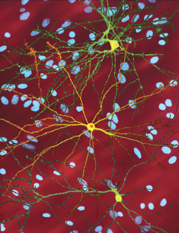
A montage of three images of single striatal neurons transfected with a disease-associated version of huntingtin, the protein that causes Huntington's disease. Nuclei of untransfected neurons are seen in the background (blue). The neuron in the center (yellow) contains an abnormal intracellular accumulation of huntingtin called an inclusion body (orange). Studies using an automated microscope and survival analysis demonstrated that neurons with disease-associated huntingtin that form inclusion bodies survive longer than those that do not.
Copyright status
This image is public domain because it was published by the National Institutes of Health
Source
File history
Click on a date/time to view the file as it appeared at that time.
| Date/Time | Thumbnail | Dimensions | User | Comment | |
|---|---|---|---|---|---|
| current | 22:55, 4 October 2007 | | 362 × 470 (60 KB) | Tian-yi (talk | contribs) | A montage of three images of single striatal neurons transfected with a disease-associated version of huntingtin, the protein that causes Huntington's disease. Nuclei of untransfected neurons are seen in the background (blue). The neuron in the center (ye |
| 22:42, 4 October 2007 |  | 362 × 470 (60 KB) | Tian-yi (talk | contribs) | A montage of three images of single striatal neurons transfected with a disease-associated version of huntingtin, the protein that causes Huntington's disease. Nuclei of untransfected neurons are seen in the background (blue). The neuron in the center (ye |
You cannot overwrite this file.
File usage
There are no pages that use this file.