File:Entamoeba trophozoites.jpg
Entamoeba_trophozoites.jpg (300 × 300 pixels, file size: 20 KB, MIME type: image/jpeg)
Summary
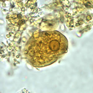
E. histolytica/E. dispar trophozoite in a direct wet mount stained with iodine.
Trophozoites: Entamoeba histolytica/Entamoeba dispar trophozoites have a single nucleus, which have a centrally placed karyosome and uniformly distributed peripheral chromatin. This typical appearance of the nucleus is not always observed as some trophozoites can have nuclei with an eccentric karyosome and unevenly distributed peripheral chromatin. The cytoplasm has a granular or "ground-glass" appearance. E. histolytica/E. dispar trophozoites usually measure 15 to 20 µm (range 10 to 60 µm), tending to be more elongated in diarrheal stool.
Erythrophagocytosis (ingestion of red blood cells by the parasite) is the only morphologic characteristic that can be used to differentiate E. histolytica from the nonpathogenic E. dispar. However, erthrophagocytosis is not typically observed on stained smears of E. histolytica.
Copyright status
This image is public domain because it was first published by the Center for Disease Control.
Source
File history
Click on a date/time to view the file as it appeared at that time.
| Date/Time | Thumbnail | Dimensions | User | Comment | |
|---|---|---|---|---|---|
| current | 19:22, 27 April 2008 | | 300 × 300 (20 KB) | Ashcraft (talk | contribs) | E. histolytica/E. dispar trophozoite in a direct wet mount stained with iodine. Trophozoites: Entamoeba histolytica/Entamoeba dispar trophozoites have a single nucleus, which have a centrally placed karyosome and uniformly distributed peripheral chromati |
You cannot overwrite this file.
File usage
There are no pages that use this file.