File:Lung cilia.jpg
Original file (1,560 × 1,257 pixels, file size: 724 KB, MIME type: image/jpeg)
Summary
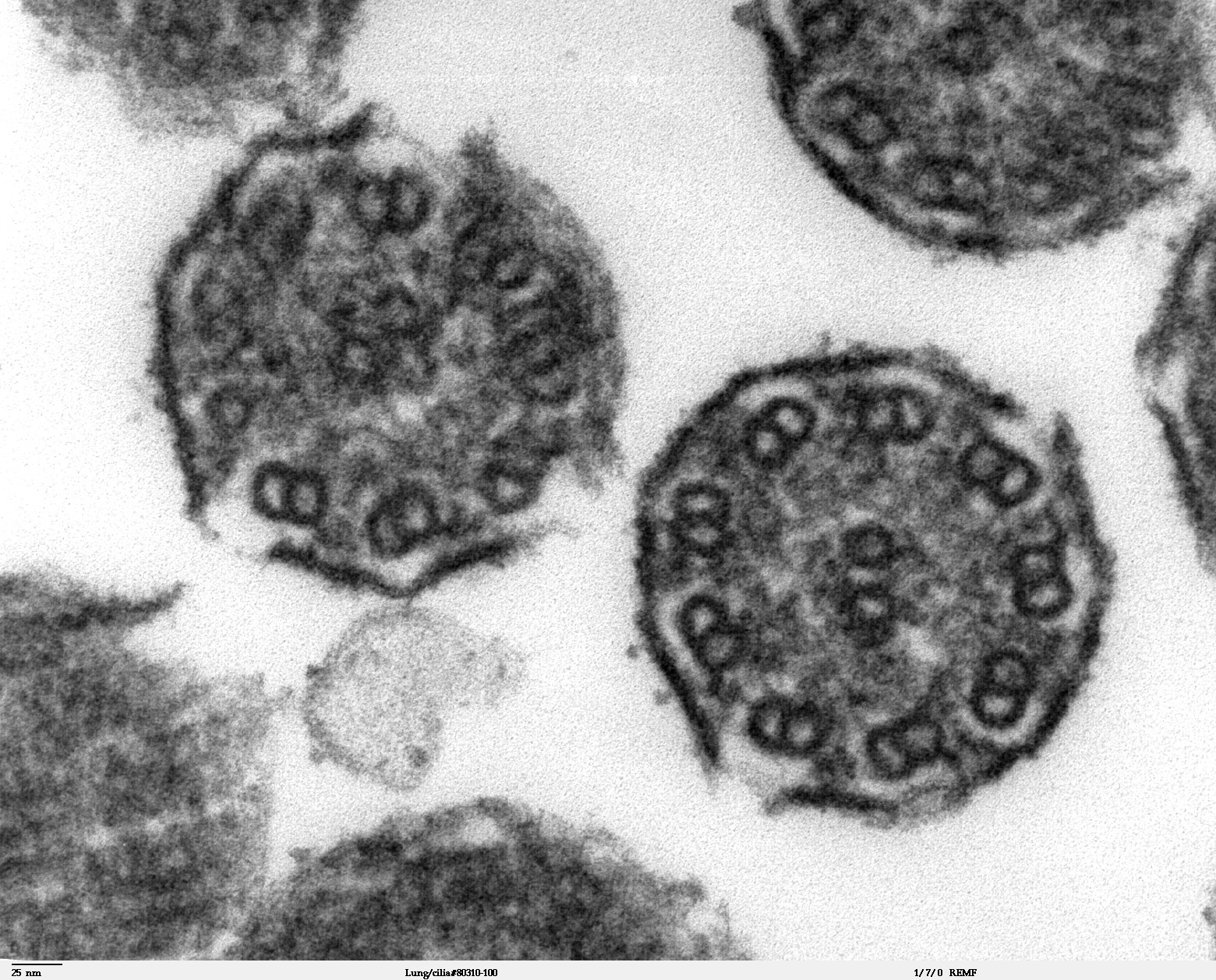
High magnification of "Lung cilia#80311-50" image. This image is a thin x-section cut through cilia. The inside of the cilium contain precisely arranged microtubles, the axoneme. The axoneme has a central unit containing two single microtubules and nine peripheral doublet microtubules (known as the "9+2"). Dynein sidearms project from the A tubule of each doublet.
Copyright status:
These images are in the public domain. Do with them what you will. If you have questions about the images or the microscopy and specimen preparation used to obtain images contact Louisa Howard . For questions regarding the research on the specimens, contact the person noted in the caption of the images. [1]
Source:
File history
Click on a date/time to view the file as it appeared at that time.
| Date/Time | Thumbnail | Dimensions | User | Comment | |
|---|---|---|---|---|---|
| current | 01:45, 28 November 2009 | | 1,560 × 1,257 (724 KB) | Ashcraft (talk | contribs) | High magnification of "Lung cilia#80311-50" image. This image is a thin x-section cut through cilia. The inside of the cilium contain precisely arranged microtubles, the axoneme. The axoneme has a central unit containing two single microtubules and nine p |
You cannot overwrite this file.
File usage
There are no pages that use this file.