File:Neuron.jpg
From CreationWiki, the encyclopedia of creation science
Jump to navigationJump to search
No higher resolution available.
Neuron.jpg (362 × 470 pixels, file size: 86 KB, MIME type: image/jpeg)
|
This image was uploaded to the shared "Image Pool" and is usable on any CreationWiki site. |
File history
Click on a date/time to view the file as it appeared at that time.
| Date/Time | Thumbnail | Dimensions | User | Comment | |
|---|---|---|---|---|---|
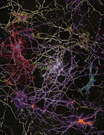
| current | 22:02, 11 October 2007 | | 362 × 470 (86 KB) | Ashcraft | A montage of four images of the development of a single neuron over a two-week period. The neuron was transfected with green fluorescent protein and an automated microscope was used to image the neuron 3 h after transfection (pseudocolored blue-green) and |
File usage
The following page uses this file: