Ficheiro:Lung cilia.jpg
De CriaçãoWiki, a enciclopédia da ciência da criação.
Saltar para a navegaçãoSaltar para a pesquisa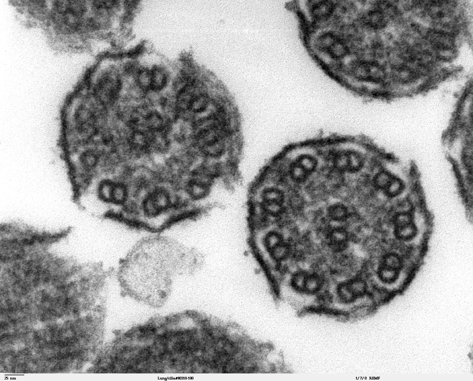
Dimensões desta antevisão: 744 × 599 píxeis. Outras resoluções: 298 × 240 píxeis | 1 560 × 1 257 píxeis.
Ficheiro original (1 560 × 1 257 píxeis, tamanho: 724 kB, tipo MIME: image/jpeg)
Histórico do ficheiro
Clique uma data e hora para ver o ficheiro tal como ele se encontrava nessa altura.
| Data e hora | Miniatura | Dimensões | Utilizador | Comentário | |
|---|---|---|---|---|---|
| atual | 01h45min de 28 de novembro de 2009 | | 1 560 × 1 257 (724 kB) | Ashcraft | High magnification of "Lung cilia#80311-50" image. This image is a thin x-section cut through cilia. The inside of the cilium contain precisely arranged microtubles, the axoneme. The axoneme has a central unit containing two single microtubules and nine p |
Utilização local do ficheiro
As seguintes 2 páginas usam este ficheiro: